
Jumat, 20 Agustus 2010
new naruto simpuuden 174
http://www.realitylapse.com/downloads/videos/naruto-shippuuden/naruto-shippuuden174-quickrelease.rmvb
Sabtu, 07 Agustus 2010
Canine distemper
Canine distemper is a very serious viral disease that affects animals in the families Canidae, Mustelidae, Mephitidae, Hyaenidae, Ailuridae, Procyonidae, Pinnipedia, some Viverridae and Felidae (though not domestic cats; feline distemper or panleukopenia is a different virus exclusive to cats). It is most commonly associated with domestic animals such as dogs, although ferrets are also vaccinated for it. It is a single-stranded RNA virus of the family paramyxovirus, and thus a close relative of measles and rinderpest.Despite extensive vaccination in many regions, it remains a major disease of dogs
Etymology
The origin of the word distemper is from the Middle English distemperen, meaning to upset the balance of the humors, which is from the Old French destemprer, meaning to disturb, which is from the Vulgar Latin distemperare: Latin dis- and Latin temperare, meaning to not mix properly
HistoryAlthough very similar to the measles virus, CDV seems to have appeared more recently, with the first case described in 1905 by French veterinarian Henri Carré. It was first thought to be related to the Plague and Typhus and resulted from several species of bacteria. It now affects all populations of domestic dog and some populations of wildlife. A vaccine was developed in 1950, yet due to limited use the virus remains prevalent in many populations. The domestic dog has largely been responsible for introducing canine distemper to previously unexposed wildlife and now causes a serious conservation threat to many species of carnivores and some species of marsupials. The virus contributed to the near-extinction of the black-footed ferret. It also may have played a considerable role in the extinction of the Tasmanian tiger and recurrently causes mortality among African wild dogs In 1991, the lion population in Serengeti, Tanzania experienced a 20% decline as a result of the disease. The disease has also mutated to form phocid distemper virus, which affects seals.
Infection
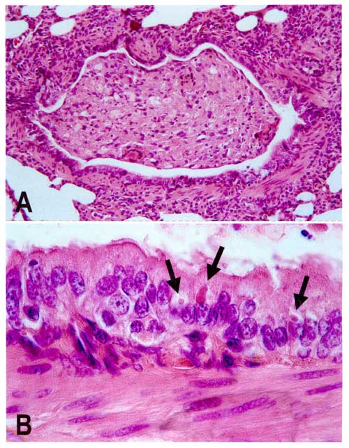
A. Lung lesion in an African Wild Dog B. Viral inclusion bodiesPuppies from three to six months old are particularly susceptible. Canine distemper virus (CDV) spreads through the aerosol droplets and through contact with infected bodily fluids including nasal and ocular secretions, feces, and urine 6–22 days after exposure. It can also be spread by food and water contaminated with these fluids. The time between infection and disease is 14 to 18 days, although there can be a fever from three to six days postinfection.
Canine distemper virus tends to orient its infection towards the lymphoid, epithelial, and nervous tissues. The virus initially replicates in the lymphatic tissue of the respiratory tract. The virus then enters the blood stream and infects the lymphatic tissue followed by respiratory, Gastrointestinal, urogenital epithelium, the Central Nervous System, and optic nerves. Therefore, the typical pathologic features of canine distemper include lymphoid depletion (causing immunosuppression and leading to secondary infections), interstitial pneumonia, encephalitis with demyelination, and hyperkeratosis of foot pads.
The mortality rate of the virus largely depends on the immune status of the infected dogs. Puppies experience the highest mortality rate where complications such as pneumonia and encephalitis are more common. In older dogs that do develop distemper encephalomyetilis, vestibular disease may present. Around 15% of canine inflammatory central nervous system diseases are a result of CDV
Disease progression
The virus first appears in bronchial lymph nodes and tonsils two days after exposure. The virus then enters the blood stream on the second or third day. In older dogs that do develop distemper encephalomyetilis, vestibular disease may present. A first round of acute fever tends to begin around 3 to 8 days after infection which is often accompanied by a low white blood cell count, especially of lymphocytes as well as low platelet count. These signs may or may not be accompanied by anorexia, a runny nose, and discharge from the eye. This first round of fever typically recedes rapidly within 96 hours and then a second round of fever begins around the 11th or 12th day and lasts at least a week. Gastrointestinal and respiratory problems tend to follow which may become complicated with secondary bacterial infections. Inflammation of the brain and spinal cord otherwise known as encephalomyelitis is either associated with this, subsequently follows, or comes completely independent of these problems. A thickening of the footpads sometimes develops and vesicularpustular lesions on the abdomen usually develop. Neurological symptoms typically are found in the animals with thickened footpads from the virus About half of sufferers experience meningoencephalitis.
Gastrointestinal and respiratory symptomsCommonly observed signs are a runny nose, vomiting and diarrhea, dehydration, excessive salivation, coughing and/or labored breathing, loss of appetite, and weight loss. When and if the neurological symptoms develop, urination and defecation may become involuntary.
Neurological symptomsThe symptoms within the central nervous system include a localized involuntary twitching of muscles or groups of muscles, seizures often distinguished by salivation and jaw movements commonly described as “chewing gum fits,” or more appropriately as "distemper myoclonus." As the condition progresses, the seizures worsen and advance to grand mal convulsions, followed by death of the animal. The animal may also show signs of sensitivity to light, incoordination, circling, increased sensitivity to sensory stimuli such as pain or touch, and deterioration of motor capabilities. Less commonly it may lead to blindness and paralysis. The length of the systemic disease may be as short as 10 days, or the start of neurological symptoms may not come until several weeks or months later. Those few that survive usually have a small tic or twitch of varying levels of severity. With time this tic will usually diminish somewhat in its severity.
Diagnosis
The above symptoms, especially fever, respiratory signs, neurological signs, and thickened footpads found in unvaccinated dogs strongly indicate canine distemper. However, several febrile diseases match many of the symptoms of the disease and only recently has differing between canine hepatitis, herpes virus, parainfluenza and leptospirosis been possible.Thus, finding the virus by various methods in the dog's conjunctival cells gives a definitive diagnosis. In older dogs that develop distemper encephalomyetilis, diagnosis may be more difficult since many of these dogs have an adequate vaccination history
The most reliable test to confirm distemper is a Brush Border slide/smear of the bladder transitional epithelium of the inside lining from the bladder, stained with Dif-Quick. These cells will always have inclusions. Inclusions in these cells which will stain a carmine red color and be para nuclear in the cytoplasm of infected cells. About 90% of the bladder cells will be positive for inclusions in the early stages of distemper. This is good for at least the first 21 days from onset of the disease. After this point, it gets harder to detect as the disease progresses further in the stages and the physical clinical signs will become quite obvious.[citation needed]
Prevention
See also: DA2PPC Vaccine
There exist a number of vaccines against canine distemper for dogs (ATCvet code: QI07AD05 and combinations) and domestic ferrets (QI20DD01), which in many jurisdictions are mandatory for pets. The type of vaccine should be approved for the type of animal being inoculated, or else the animal could actually contract the disease from the vaccine. A dog who has eaten meat infected with Rinderpest can also sometimes receive temporary immunity.Infected animals should be quarantined from other dogs for several months due to the length of time the animal may shed the vires. The virus is destroyed in the environment by routine cleaning with disinfectants, detergents, or drying. It does not survive in the environment for more than a few hours at room temperature (20–25 °C), but can survive for a few weeks in shady environments at temperatures slightly above freezing. It, along with other labile viruses, can also persist longer in serum and tissue debris
Treatment
Until recently, canine distemper has been associated with a long history of pessimism with respect to treatment of infected animals and the disease was usually assumed to have a poor prognosis. Most care offered was only palliative, geared toward easing the suffering. Several factors had an important role in maintaining the status quo.
Misdiagnosis, miseducation, a lack of treatment, inadequate, or inappropriate treatment has historically created barriers and slowed the development of effective solutions to the disease. Even today the needs of affected animals often go unrecognized until the disease reaches the nervous stage and the distressed behavior and/or impaired functional state of the animal is more obvious and less responsive to treatment.
Research and funding for the most part have focused on preventative vaccination rather than on finding a cure for distemper.
Another factor is the outdated theory that the injuries that occurred were the result of a strictly autoimmune reaction, the thought being that initially the canine distemper virus was introduced, then subsequently eliminated, but that cytokines continued to attack and damage healthy tissue in the absence of a current pathogen. Based on that faulty assumption, anti-inflammatory and immunosuppressive drugs have been prescribed by some veterinarians in an attempt to bring the effects of the condition under control.
It was later considered that the action of macrophages on nerve cells is targeted on infected cells, indicating that the autoimmune reaction is likely a direct consequence of the presence of the virus Often, owners seek expert help only when the disease is in its advanced stages (nervous phase) and prescription anti-inflammatory drugs (which are usually corticosteroids) undermine the immune system of the animal, allow the proliferation of the virus, and the autoimmune reaction increases as a means of containment of infected cells.
The most successful treatments for canine distemper are adaptations of established treatments used for other diseases caused by similar viruses, such as Ribavirin and vitamin A, which are used to treat measles, the same family and gender (Paramyxoviridae - morbillivirus) and Interferon Alpha, used for the treatment of measles and a vaccine used to immunize birds against Newcastle disease same family but different genera (Paramyxoviridae - AVULAVIRUS).
The first references that suggested effective treatments for similar viruses could be effective for canine distemper arose when studies found that canine distemper was a disease comparable to measles and infected animals could be used to develop new technologies for treatment of measles. The question of whether the reciprocal would be true was cleared when studies assessed the efficacy of traditional treatments for measles which were then successfully applied to animals with distemper.
The first finding was when the induction of high levels of Vitamin A, which is a ostensibly treatment used to treat measles (including being recommended by the World Health Organization), produced an effect of 100% cure in animals experimentally infected. The group that received no supplementation all died. . Currently, it is known that the direct inhibitor effect of retinoids (Vitamin A and subproducts) over the measles virus is what confirms the choice of Vitamin A as a treatment for canine distemper
The confirmation of the effectiveness of Vitamin A in the treatment of canines, especially dogs, is its ability to convert the Vitamin A into nontoxic esters This characteristic of carnivores is well known, which removes the risk of hypervitaminosis possible due to the maintenance of high doses. For dogs there is a benchmark to measure the risk of hypervitaminosis, a national research found that it takes a dose of 300,000 IU / kg daily for thirty days so that the first signs of hypervitaminosis appear, and need sixty days of ingestion at this dosage to kill the animal Since this dosage, 300,000 IU / kg is sixty times greater than the limit established for humans.
The mechanisms of action that explain its effectiveness in the treatment of distemper remain unexplained, and this issue also exists for the case of measles. Some evidence points to an indirect action, such as checking that there is a reduction in the amounts of Vitamin A during infection, pointing to the hypothesis that [ [Vitamin A]] is raw material for some mechanism of resistance to infection. The very characteristic antiinfectiva not specific Vitamin A is a mystery, however there was any doubt about its effectiveness, action mechanisms elucidated or not.
The adoption of Ribavirin as a treatment for canine followed the same steps of Vitamin A, it was the principle used in cases of subacute sclerosing panencephalitis under measles. The first verification of the effectiveness occurred in vitro. What was observed was that the distemper virus is very susceptible to Ribavirin and its mechanism of induction error catastrophe are needed from 0.02 to 0.05 micro mols to create an inhibitory effect on virus replication by 50%.
The main concern in the use of Ribavirin was the result of its interaction with the blood-brain barrier. Being the brain a immunologically privileged area, the concerns was the capacity of Ribavirin to overcome this barrier. A study using mice with encephalitis due to measles it was found that once the virus has become established in phase nervosa, the blood-brain barrier in a way fails, reducing the restriction to the action of the Ribavirin in these areas. The verification of all these results in vivo resulted in an effectiveness of 80% in animals that had already reached the nervous phase of viral infection. The application of Ribavirin demands a close monitoring of the animal due the risk of leukopenia and the ingestion of log-chain tryglicerides(in other words: fats) in order to better absorb the drug and for preservation of gastric region, which are quite susceptible to Ribavirin.[dubious – discuss]
Canine distemper virus and Paget's disease
Paget's disease, a focal destructive disease of bone, has long suspected paramyxoviruses such as CDV, measles, respiratory syncytial virus, simian virus 5, and parainfluenza virus Type 3 as a culprit. Most studies, however, have pointed more directly at CDV and Measles.[33][34][35] The virus detection technique in situ-RT-PCR has shown CDV in 100% of Pagetic samples whereas other virus detection techniques have been less accurate
Etymology
The origin of the word distemper is from the Middle English distemperen, meaning to upset the balance of the humors, which is from the Old French destemprer, meaning to disturb, which is from the Vulgar Latin distemperare: Latin dis- and Latin temperare, meaning to not mix properly
HistoryAlthough very similar to the measles virus, CDV seems to have appeared more recently, with the first case described in 1905 by French veterinarian Henri Carré. It was first thought to be related to the Plague and Typhus and resulted from several species of bacteria. It now affects all populations of domestic dog and some populations of wildlife. A vaccine was developed in 1950, yet due to limited use the virus remains prevalent in many populations. The domestic dog has largely been responsible for introducing canine distemper to previously unexposed wildlife and now causes a serious conservation threat to many species of carnivores and some species of marsupials. The virus contributed to the near-extinction of the black-footed ferret. It also may have played a considerable role in the extinction of the Tasmanian tiger and recurrently causes mortality among African wild dogs In 1991, the lion population in Serengeti, Tanzania experienced a 20% decline as a result of the disease. The disease has also mutated to form phocid distemper virus, which affects seals.
Infection
A. Lung lesion in an African Wild Dog B. Viral inclusion bodiesPuppies from three to six months old are particularly susceptible. Canine distemper virus (CDV) spreads through the aerosol droplets and through contact with infected bodily fluids including nasal and ocular secretions, feces, and urine 6–22 days after exposure. It can also be spread by food and water contaminated with these fluids. The time between infection and disease is 14 to 18 days, although there can be a fever from three to six days postinfection.
Canine distemper virus tends to orient its infection towards the lymphoid, epithelial, and nervous tissues. The virus initially replicates in the lymphatic tissue of the respiratory tract. The virus then enters the blood stream and infects the lymphatic tissue followed by respiratory, Gastrointestinal, urogenital epithelium, the Central Nervous System, and optic nerves. Therefore, the typical pathologic features of canine distemper include lymphoid depletion (causing immunosuppression and leading to secondary infections), interstitial pneumonia, encephalitis with demyelination, and hyperkeratosis of foot pads.
The mortality rate of the virus largely depends on the immune status of the infected dogs. Puppies experience the highest mortality rate where complications such as pneumonia and encephalitis are more common. In older dogs that do develop distemper encephalomyetilis, vestibular disease may present. Around 15% of canine inflammatory central nervous system diseases are a result of CDV
Disease progression
The virus first appears in bronchial lymph nodes and tonsils two days after exposure. The virus then enters the blood stream on the second or third day. In older dogs that do develop distemper encephalomyetilis, vestibular disease may present. A first round of acute fever tends to begin around 3 to 8 days after infection which is often accompanied by a low white blood cell count, especially of lymphocytes as well as low platelet count. These signs may or may not be accompanied by anorexia, a runny nose, and discharge from the eye. This first round of fever typically recedes rapidly within 96 hours and then a second round of fever begins around the 11th or 12th day and lasts at least a week. Gastrointestinal and respiratory problems tend to follow which may become complicated with secondary bacterial infections. Inflammation of the brain and spinal cord otherwise known as encephalomyelitis is either associated with this, subsequently follows, or comes completely independent of these problems. A thickening of the footpads sometimes develops and vesicularpustular lesions on the abdomen usually develop. Neurological symptoms typically are found in the animals with thickened footpads from the virus About half of sufferers experience meningoencephalitis.
Gastrointestinal and respiratory symptomsCommonly observed signs are a runny nose, vomiting and diarrhea, dehydration, excessive salivation, coughing and/or labored breathing, loss of appetite, and weight loss. When and if the neurological symptoms develop, urination and defecation may become involuntary.
Neurological symptomsThe symptoms within the central nervous system include a localized involuntary twitching of muscles or groups of muscles, seizures often distinguished by salivation and jaw movements commonly described as “chewing gum fits,” or more appropriately as "distemper myoclonus." As the condition progresses, the seizures worsen and advance to grand mal convulsions, followed by death of the animal. The animal may also show signs of sensitivity to light, incoordination, circling, increased sensitivity to sensory stimuli such as pain or touch, and deterioration of motor capabilities. Less commonly it may lead to blindness and paralysis. The length of the systemic disease may be as short as 10 days, or the start of neurological symptoms may not come until several weeks or months later. Those few that survive usually have a small tic or twitch of varying levels of severity. With time this tic will usually diminish somewhat in its severity.
Diagnosis
The above symptoms, especially fever, respiratory signs, neurological signs, and thickened footpads found in unvaccinated dogs strongly indicate canine distemper. However, several febrile diseases match many of the symptoms of the disease and only recently has differing between canine hepatitis, herpes virus, parainfluenza and leptospirosis been possible.Thus, finding the virus by various methods in the dog's conjunctival cells gives a definitive diagnosis. In older dogs that develop distemper encephalomyetilis, diagnosis may be more difficult since many of these dogs have an adequate vaccination history
The most reliable test to confirm distemper is a Brush Border slide/smear of the bladder transitional epithelium of the inside lining from the bladder, stained with Dif-Quick. These cells will always have inclusions. Inclusions in these cells which will stain a carmine red color and be para nuclear in the cytoplasm of infected cells. About 90% of the bladder cells will be positive for inclusions in the early stages of distemper. This is good for at least the first 21 days from onset of the disease. After this point, it gets harder to detect as the disease progresses further in the stages and the physical clinical signs will become quite obvious.[citation needed]
Prevention
See also: DA2PPC Vaccine
There exist a number of vaccines against canine distemper for dogs (ATCvet code: QI07AD05 and combinations) and domestic ferrets (QI20DD01), which in many jurisdictions are mandatory for pets. The type of vaccine should be approved for the type of animal being inoculated, or else the animal could actually contract the disease from the vaccine. A dog who has eaten meat infected with Rinderpest can also sometimes receive temporary immunity.Infected animals should be quarantined from other dogs for several months due to the length of time the animal may shed the vires. The virus is destroyed in the environment by routine cleaning with disinfectants, detergents, or drying. It does not survive in the environment for more than a few hours at room temperature (20–25 °C), but can survive for a few weeks in shady environments at temperatures slightly above freezing. It, along with other labile viruses, can also persist longer in serum and tissue debris
Treatment
Until recently, canine distemper has been associated with a long history of pessimism with respect to treatment of infected animals and the disease was usually assumed to have a poor prognosis. Most care offered was only palliative, geared toward easing the suffering. Several factors had an important role in maintaining the status quo.
Misdiagnosis, miseducation, a lack of treatment, inadequate, or inappropriate treatment has historically created barriers and slowed the development of effective solutions to the disease. Even today the needs of affected animals often go unrecognized until the disease reaches the nervous stage and the distressed behavior and/or impaired functional state of the animal is more obvious and less responsive to treatment.
Research and funding for the most part have focused on preventative vaccination rather than on finding a cure for distemper.
Another factor is the outdated theory that the injuries that occurred were the result of a strictly autoimmune reaction, the thought being that initially the canine distemper virus was introduced, then subsequently eliminated, but that cytokines continued to attack and damage healthy tissue in the absence of a current pathogen. Based on that faulty assumption, anti-inflammatory and immunosuppressive drugs have been prescribed by some veterinarians in an attempt to bring the effects of the condition under control.
It was later considered that the action of macrophages on nerve cells is targeted on infected cells, indicating that the autoimmune reaction is likely a direct consequence of the presence of the virus Often, owners seek expert help only when the disease is in its advanced stages (nervous phase) and prescription anti-inflammatory drugs (which are usually corticosteroids) undermine the immune system of the animal, allow the proliferation of the virus, and the autoimmune reaction increases as a means of containment of infected cells.
The most successful treatments for canine distemper are adaptations of established treatments used for other diseases caused by similar viruses, such as Ribavirin and vitamin A, which are used to treat measles, the same family and gender (Paramyxoviridae - morbillivirus) and Interferon Alpha, used for the treatment of measles and a vaccine used to immunize birds against Newcastle disease same family but different genera (Paramyxoviridae - AVULAVIRUS).
The first references that suggested effective treatments for similar viruses could be effective for canine distemper arose when studies found that canine distemper was a disease comparable to measles and infected animals could be used to develop new technologies for treatment of measles. The question of whether the reciprocal would be true was cleared when studies assessed the efficacy of traditional treatments for measles which were then successfully applied to animals with distemper.
The first finding was when the induction of high levels of Vitamin A, which is a ostensibly treatment used to treat measles (including being recommended by the World Health Organization), produced an effect of 100% cure in animals experimentally infected. The group that received no supplementation all died. . Currently, it is known that the direct inhibitor effect of retinoids (Vitamin A and subproducts) over the measles virus is what confirms the choice of Vitamin A as a treatment for canine distemper
The confirmation of the effectiveness of Vitamin A in the treatment of canines, especially dogs, is its ability to convert the Vitamin A into nontoxic esters This characteristic of carnivores is well known, which removes the risk of hypervitaminosis possible due to the maintenance of high doses. For dogs there is a benchmark to measure the risk of hypervitaminosis, a national research found that it takes a dose of 300,000 IU / kg daily for thirty days so that the first signs of hypervitaminosis appear, and need sixty days of ingestion at this dosage to kill the animal Since this dosage, 300,000 IU / kg is sixty times greater than the limit established for humans.
The mechanisms of action that explain its effectiveness in the treatment of distemper remain unexplained, and this issue also exists for the case of measles. Some evidence points to an indirect action, such as checking that there is a reduction in the amounts of Vitamin A during infection, pointing to the hypothesis that [ [Vitamin A]] is raw material for some mechanism of resistance to infection. The very characteristic antiinfectiva not specific Vitamin A is a mystery, however there was any doubt about its effectiveness, action mechanisms elucidated or not.
The adoption of Ribavirin as a treatment for canine followed the same steps of Vitamin A, it was the principle used in cases of subacute sclerosing panencephalitis under measles. The first verification of the effectiveness occurred in vitro. What was observed was that the distemper virus is very susceptible to Ribavirin and its mechanism of induction error catastrophe are needed from 0.02 to 0.05 micro mols to create an inhibitory effect on virus replication by 50%.
The main concern in the use of Ribavirin was the result of its interaction with the blood-brain barrier. Being the brain a immunologically privileged area, the concerns was the capacity of Ribavirin to overcome this barrier. A study using mice with encephalitis due to measles it was found that once the virus has become established in phase nervosa, the blood-brain barrier in a way fails, reducing the restriction to the action of the Ribavirin in these areas. The verification of all these results in vivo resulted in an effectiveness of 80% in animals that had already reached the nervous phase of viral infection. The application of Ribavirin demands a close monitoring of the animal due the risk of leukopenia and the ingestion of log-chain tryglicerides(in other words: fats) in order to better absorb the drug and for preservation of gastric region, which are quite susceptible to Ribavirin.[dubious – discuss]
Canine distemper virus and Paget's disease
Paget's disease, a focal destructive disease of bone, has long suspected paramyxoviruses such as CDV, measles, respiratory syncytial virus, simian virus 5, and parainfluenza virus Type 3 as a culprit. Most studies, however, have pointed more directly at CDV and Measles.[33][34][35] The virus detection technique in situ-RT-PCR has shown CDV in 100% of Pagetic samples whereas other virus detection techniques have been less accurate
Parvovirus
WHAT IS PARVOVIRUS?

Canine Parvovirus is a viral disease of dogs that was first reported in early 1978. Parvovirus is capable of causing two different sets of clinical problems. The first to be recognized, and most common, is the “intestinal” form, which is manifested by diarrhea; often bloody vomiting, loss of appetite, depression, fever, and sometimes death. The second syndrome, the “cardiac” form, occurs in very young pups and is manifested by an acute inflammation of the heart muscle.
Any age, breed or sex of dog could be affected by Parvovirus. However, infection with Parvovirus does not automatically mean illness. Several factors such as age, environment, stress, parasites and general health status of each individual dog infected could affect the severity of illness. The degree of illness could range from very mild to unapparent to very severe, often resulting in death. The disease is usually more severe in young dogs (less than 6 months of age) or old dogs. Doberman pinschers and Rottweilers are breeds that are more susceptible to parvovirus.
WHERE DID IT COME FROM?
Experts agree that canine Parvovirus is closely related to Parvoviruses that affect other animals. Where the virus originally came from remains unknown, but it is possible that it is a mutant from another Parvovirus that affects other species of animals. Man is not known to be affected by canine Parvovirus. Since its first appearance in 1978, canine Parvovirus has spread to every continent in the world, probably the result of the hardy nature of the virus. It is resistant to extremes of temperature (i.e., it survives freezing and extreme heat) and is unharmed by detergents, alcohols and common disinfectants. Direct transmission occurs when an infected dog comes in contact with a healthy dog. The virus is found in heavy concentration in the infected dog’s stool. The virus particles can be easily spread on shoes, clothing and other inanimate objects. Fleas, as well as people, can therefore act as indirect sources of infection.
Oral Phase:
The disease process begins with the oral ingestion of Parvovirus from the feces of an infected dog. The virus initially invades the lymph glands of the throat (lymph nodes and tonsils) where it multiplies. Following multiplication in the lymph glands for l to 2 days, the virus then enters the blood stream, which causes the VIREMIA phase (virus in the blood).

Viremia Phase:
This phase is characterized by massive amounts of virus in the dog’s bloodstream, which in turn is spread to all parts of the body, such as, the intestine, bone marrow, spleen, other lymph nodes and the heart (in young pups less than 8 weeks of age). As infection spreads, the symptoms of illness become apparent. (See “symptoms” in next section). The Viremia phase can last for approximately l to 9 days.
Contagious Stage:
The final phase in the cycle is the contagious or “shedding” phase. As many as 30 billion Parvovirus particles can be shed from the intestines of an infected dog in every ounce of stool. The highest concentration of virus in the stool is seen when the infected dog is showing signs of illness. A dog can, however, be a source of infection to other dogs without having observable signs of illness. Transmission can occur for at least 3 weeks after a dog becomes infected with the virus. Chronic “carriers” are not know to exist as in other virus disease. Parvovirus in the environment can infect susceptible dogs for many months once shed in the stool.
WHAT ARE THE SYMPTOMS?
The symptoms below indicate a problem warranting medical attention. Early, vigorous treatment of illness caused by canine Parvovirus infection is imperative since vomiting and diarrhea can lead to dehydration and chemical imbalance in the body. If your dog shows these signs, see your veterinarian. Early treatment can save lives.
Cardiac Form
- Sudden death
- Crying, difficulty breathing, gasping for breath
- Extreme depression
- Weakness
- Unwillingness to nurse
- Irregular heartbeat
Intestinal Form (any age dog affected, but more severe in puppies).
- Depression
- Loss of appetite
- Fever (above 103 degrees F)
- Vomiting
- Diarrhea with or without blood (more serious if blood present)
- Low white blood count
HOW IS IT CONTROLLED?
Control of Parvovirus by sanitation measures alone is extremely difficult because the virus is such a resistant, hardy organism and because it is so easily spread. Contact with other dogs and especially their stool, should be minimized. Clorox diluted one part to 30 parts with water has been effective in disinfecting inanimate objects such as clothing, floors, kennels, etc. However, it is impractical, if not impossible, to disinfect public streets, parks, etc. Isolation of infected dogs is another method of control, although moderately effective. Both of these measures will help reduce the amount of contagious virus in the environment, but only vaccination will control the actual source of infection, the contagious shedding dog.

Vaccination:
The most effective control measure for canine Parvovirus disease. A properly immunized dog will have circulating antibodies in the blood that will destroy Parvovirus following exposure.
THE ROLE OF MATERNAL ANTIBODY IN PUPPY IMMUNIZATION:
Maternal antibodies are antibodies against Parvovirus, which are passed from the mother to the puppies through the “first milk” or colostrum. They provide the puppy with an immediate temporary or “passive” immunity. The mother obtains these antibodies from prior vaccination or by natural exposure to Parvovirus. However, maternal antibody is a two-edged sword; it protects the puppy against disease early in life, but it also blocks active immunization. In the case of Parvovirus, maternal antibody can interfere with vaccination for as long as 14 to 16 weeks of age in some pups. A refractory period can exist in some pups where very low, almost undetectable levels of maternal antibody will inhibit the vaccination process but will not prevent Parvovirus infection. Since the level of maternal antibody varies from puppy to puppy, it is important to begin vaccination at an early age and repeat every 2-3 weeks until the puppy is at least 16 - 18 weeks old.


Struktur sel Bakteri
Bakteri sebagai organisme prokariotik yang merupakan organisme uniseluler memiliki struktur sel yang tidak memiliki membran inti. Struktur sel secara umum yang dimiliki oleh sel prokariot dapat kita lihat pada sel bakteri.

Struktur Sel Bakteri
Nukleoid (Nukleus).Nukleus atau inti sel berfungsi sebagai pengendali dan pengatur sel. seluruh aktifitas sel diatur oleh nukleus. Nukleus juga berfungsi sebagai pembawa informasi genetik yaitu kromosom, yang diwariskan ke generasi selanjutnya. Kromosom adalah struktur yang tersusun oleh molekul DNA dan protein (histon). Nukleus sel bakteri terpapar atau kontak langsung dengan sitoplasma karena tidak memiliki membran inti.
Cytoplasm (Sitoplasma). Sitoplasma adalah bagian sel yang berisi cairan tempat berlangsungnya metabolisme sel. Kandungan terbesar dalam sitoplasma adalah air (80-90%).
Ribosome (Ribosom). Ribosom merupakan struktur berupa butiran-butiran kecil yaang merupakan tempat sintesis protein. Protein disintesis atau dibuat dengan menggabungkan beberapa asam amino yang sesuai informasi genetik yang ada di molekul DNA. Ribosom berada di sitoplasma.
Cytoplasmic membrane (Membran Plasma). Membran plasma adalah lapisan di luar sitoplasma yang tersusun atas phospolipid bilayer. Fungsi membran plasma adalah sebagai pelindung dan mengatur transportasi sel. Pengaturan transportasi sel dimasksudkan untuk mengatur keluar masuknya substansi ke dalam dan ke luar sel. Membran plasma juga berperan dalam penerima rangsang yang datang dari luar sel.
Membran sel pada sel prokariot mengalami pelekukan ke arah dalam membentuk struktur yang disebut mesosome (mesosom). Mesosom berfungsi sebagai tempat terjadinya respirasi sel sehingga dihasilkan energi yang akan digunakan untuk aktifitas di dalam sel.
Cell wall (Dinding Sel). Dinding sel adalah struktur pelindung kedua setelah membran plasma.
Cell wall (Dinding Sel). Dinding sel adalah struktur pelindung kedua setelah membran plasma.
Capsule (Kapsul). Kapsul adalah struktur pelindung sel ketiga setelah membran plasma dan dinding sel.
Pili (Bulu Rambut). Pili berfungsi sebagai alat pelekatan sel bakteri pada suatu permukaan substrat atau benda. Flagella (Flagel). Flagel berfungsi dalam pergerakan sel. Baik flagel dan pili disusun oleh mikrotubulus.
Jumat, 30 Juli 2010
muhasabah mau ramadhan
Sesungguhnya seorang hamba jika melakukan dosa, maka terbentuklah noda hitam dalam hatinya. Jika ia melepaskan dosa, istighfar dan taubat, bersihlah hatinya. Ketika mengulangi dosa lagi, bertambahlah noda hitamnya, sehingga menguasai hati. Itulah Roon (rona) yang disebutkan dalam Al-Qur’an, “Sekali-kali tidak (demikian), Sebenarnya apa yang selalu mereka usahakan itu menutupi hati mereka.” (HR At-Tirmidzi).
Selasa, 23 Maret 2010

Sabtu, 06 Maret 2010
PK
PATOLOGI KLINIK
PATOLOGI : mempelajari gangguan struktur suatu organ.
PATOLOGI KLINIK : Mempelajari gangguan fungsi fisiologi suatu organ atau jaringan berdsarkan hasil lab untuk kepentingan klinik (prognosa, diagnosa, terapi)
Tujuan Patologi klinik :
1. Membantu mendiagnosa penyebab penyakit
2. Mengetahui kondisi hewan untuk kepentingan terapi.
3. Mangetahui prognosa.
• Pemeriksaan Darah .
• Leukositosis (meningkat) karena bakteri (paru2)
• Leukopenia (menurun) karena virus (gambaran virus)
• Deferensial lukosit seperti;
o netrofil karena Bakteri
o eosinofil karena parasit
o limfosit karena Virus
semua itu bertujuan untuk diagnosa
• Total HG, eritrosit dan trombosit untuk tujuan terapi.
Hematokrit meningkat adalah dehidrasi (type; ringan,, tepi ada, berat), hematokrit 6% tempati darah. Anemia apa? Normositik yaitu pendarahan (darah keluar dari pembuluh darah), berdilitasi oleh vitamin2 terutama vitamin K. Akut kronis biasanya karena habis divaksin dijaringan lihat makrofag. MCV yaitu ukuran eritrosit. Makrositik : fak hemopoisis(tidak bisa dewasa) , defesiensi vitamin B12, B6, aasam folat. Mikrositik : defesiaensi fe, parasit kronis. MCHC : warna eritrosit ditentukan oleh haemoglobin. Hipokromik : HG menurun dan Fe menurun, Hiperkramik : HG meningkat (digabung2).
Pemeriksaan total trombosit : perdarahan yaitu trombosit menurun. Trombositepenia yaitu induksi perdarahan karena trombosit terpakai untuk pembekuan darah. Prognosa : infausta, fausta.
• GINJAL untuk pemeriksaan lab : urin dan darah.
Trombosit rendah karena pendarahan dan untuk terpakai pembekuan darah. Kalau ada perubahan : trombosit tumpah ke tempat luka. Fibrin akan terlihat.
• BLOOD URINE NITROGEN
Pemeriksaan BUN biasanya kelemahan pada hewan banyak makan protein BUN meningkat padahal ginjal normal.
Kreatinin (daging banyak, dehidrasi, gannguan ginjal) yang lebih sensitif karena kreatinin hasil akhir dari kreatinin otot. Kreatini meningkat akan terjadi gagal ginjal.
Daging pembentukan asam dgn amino lalu hasil akhir urea. Kondisi dehidrasi, urea diserap oleh tubulus ke pembuluh darah urea meningkat.
• SGPT lebih sensitif untuk kerusakan sel hati
• Bilirubin total dan direk untuk ikterus
• Total protein, albumin, Globulin untuk sintesis hati.
• Untuk tes hati, ginjal, lemak
o SGOT , SGPT (lebih banyak di hati) ; untuk melihat fosfat alkali, kerusakan sel hati SGPT/SGOP, alatnya reclotron.
o Untuk melihat hati ikterus pada bilirubin total / bilirubin direk
o Untuk tes hati sintesa protein : albumin, globilin dan total protein(refraktometer) BJ
o Glukosa yaitu kadar gula darah ditemukan pada diabetes militus 90/125 normal, meningkat (Glikesemenia), menurun (hypoglikeseminia)
o Tipe diabet ; 1. Pancreas gagal produksi insulin karena glukosa langsung di glikogen. 2. Pancreas maupun membuat insulin harus ada mendorong pemberian abat pendorong insulin (amerik)
Senin, 01 Maret 2010
GANGGUAN ALAT PENCERNAAN
- MULUT, PHARING, OSEPHAGUS, LAMBUNG, USUS HALUS,BESAR DAN REKTUM.
1. MULUT
- Ada benda asing
- Radang
- Kelainan gigi
- Tumor
- Salivari = beri atropin
2. PHARING
- Benda asing
- Radang (Antibiotik)
- Gak mau makan ( AB, vit), muntah
- Batuk (obat)
- Tumor (operasi)
- Paralisis (susah diobatin)
3. OSEPHAGUS
- Ocrivicalis = Palpasi
- Thorecalis = USG, Radiologi, Endocospy
- Obstruksi/ benda asing = berusaha menelan tapi tidak bisa. Gunakan Terapi Atropin (lalu di dorong ke dalam) AB sistemik. Antihelmintika sistemik.
4. LAMBUNG (tunggal)
- Dilatasi lambung
- Tukak lambung (ulcer pada lambung)
- Benda asing
- Tumor
- Parasit lambung pilucer kaligi
- Gejala : muntah (gangguan pharing), palpasi costae terakhir, gangguan hati, ginjal, pangkreas, paru2.
- Gangguan pada lambung ; ada ulcer, radang pada lambung, alergi. Gastritis : terapi Antihistamin, infeksi dengan antibiotika, sedativa lambung. Pyramid amo ductiva (prolotag).
- Terapi treatament = banyak muntahan diinfus kalau ada benda asing di rotgen
- Batuk paroximal(pharingitis)
- Ada sumbatan pada pylorus
- Terlalu penuh.
5. USUS
- Obstruksi
- Valvolasi = muntah persisten, dehidrasi (pcesesti = perbaiki usus) terapi operasi
- Stragulasi
- Kalau terjadi gangguan usus halus dan besar yaitu feces apa ada parasit, protozoa dan sebagainya.
- Gangguan di usus = ada peningkatan motilitas ( otot lelah = constipasi), penurunan motilitas
- Penyebab meningkat/menurun mortilitas dikarenakan alergi ( ganagguan motilitas)
- Kepekaan meningkat (radang) maka peristaltiknya akana meningkat.
- Terapi = menaikan/menurunkan kalor air. Atropin ( sinaps impuls tidak bisa jalan) antregensin ( reseptornya di blok). Misalnya naik maka diturunkan dengan meningkatkan peristaltiknya dengan menggunakan obat yang menurunkana syaraf sedativa usus untuk mengobati rangsangan dengan meningkatkan peristaltik kemudian diberikan obat parositolitik (berpengaruh pada syaraf otonom) terutama pada jantung (hati – hati cek jantung)
- Pengobatan symtomatis = kalau ada parasit gunakan antihelmint, bakteri dengan antibiotika, alergi dengan antihistamin.
- Infeksi bisa disebabkan karena alergi kemudian berikan antihistamin dan antibiotika. Dengan menurunkan motilisas dengan furgativa = syaraf furgativa. Osmotik furgativa ( garam inggris) menyerap cairan extra seluler di usus.
- Pada colon (triccicis cubi) = hampir sama rangsangan buang air lebih tinggi, obat2 sama, vitamin larut dalaam air B . Complek.
- Rectum kejadian sering terjadi konstipasi = coprotasis ( dilihat di anjing bertubuh panjang). Pulsus kontipasi karena bulu2 pada Pseudococustasis(bulu hilangin, feces dikeluarin) dari pihak saatu terhalangi atau dihalangi untuk di enema (masukan cairan), gliserin, sabun; setelah keluar diberikan lantaxia dengan gerakan peristaltik dengan glukolale.
- Pada anus glandula sirconane karena gatal, benjolan2 ( radang nanahnya dikeluarin dengan punctio) diobati, sistemik, injeksi.
6. HEPAR
- Pemeriksaan secara (palpasi kanan coste terakhir) radang akut bisa dillihat.
- Infeksi = muntah, jundis pada mukosa (konsentrasi empedu kekuningan) hepatik, prehepatik. ( empedu dari eritrosit pecah kemudian fe ke bilirubin kemudian globin(protein). ( indirec bilirubin = pecah dari bilirubin)
- Metabolisme eritrosit oleh parasit.
- Hepatik menjadi proses empedu tidak di proses (kerusakan pada hati) empedu beredar dalam darah. Empedu masuk ke hati di konjugasikan, colirunat)
- Post hepatik = bilirubin di konjugasikan masuk ke dalam empedu, karen terjadi obstruksi pada saluran empedu( sehingga tidak bisa keluar.
- Empedu yang masuk ke usus di reduksi menjadi urobirinogen ada yang di serap kembali oleh vena vorta dan sekresi olehh ginjal.
- Bilirubin tidak ada di urin menjadi berarti ada obstruksi di hati
- Kalau dilihat di hepaatik, prehepatik, post hepatik lihat jundice di indexnya(kucing2) kalau lebih ada gangguan.
- Penekanan dengan lab.
- Gangguan hati = sintesa protein (globulin) hipoglobunia = ada gangguan di hati (infeksi)
- Protombin sebagai bahan pembeku darah, vitamin k, larut dalam lemak.
- Gangguan metabolisme lemak menjadi gangguan vitamin k kemudian protombin terganggu coal gutin time di perpanjang.
- Kolesrerol tinggi karena di bentuk dalam jaringan kemudian dirubah dalam hati oleh asam folat. Kalau hati rusak perubahan itu terjadi sehingga kolesterol tinggi.
- Asam urat, urid acid karena dibentuk oleh purin (kacang2n dan lemak = jeroan) purin dalam hati dirubah menjadi asam urat dirubah dalam hati = sehingga asam urat tinggi.
- SGOT & SGPT. SGOT juga di produksi dalam jaringan termasuk dalam hati kalau hati rusak SGOT tinggi. SGPT konsentrasinya lebih tinggi di hati karena kalau rusakan HEPATOSELULER.
- Kalau rusak 70 % regenerasinya tinggi (baru kelihatan gejala)
- Pengobatan = infeksi = hepatitis, virus, bakteri = antibiotika . hepar ada muntah terapi dymtomatis, pemeliharaan sel hati karena metilin dan urin, ada parasit = flagil / subculamin(toksik), biasa gangguan acites = cairan di functio = diuretikan = flusamix, lasix.
7. PANKREAS
- Endokrin ; insulin ( menurunkan kadar gula darah), glukagon ( bekerja menaikan kadar gula darah)
- Exsokrin ; lipase(membantu getah2 pencernaan) lemak, Amilase(karbohidrat), tripsin(protein)
- Tanda2 klinis kalau akut = terjadi rasa sakit amat sangat colic palsu.
- Comfirmasi di feces = ada lemak2, serat2 daging utuh, yang terjadi diare (lasansia)
- Ada gangguan di pangkreas= oposisinya = karena obesitas, asam lemak tinggi mengganggu metabolisme tersebut.
- Gangguan pada pangkreas, sistem eksokrim di bantu oleh luar sama dengan diabet. Enzim pancreas = pancreatin. Kalau ada radang = antibiotika(diare persisten) karena terjadi amilase meningkat lipase dan uremia keadaan ini hampir sama dengan gangguan ginjal. Test vitamin A, (larut dalam lemak) karena terganggu dalam darah (perlu alat khusus)).
- Gaangguan endokrin = insulin = kadar gula darah di periksa kalau lebih dari 100 (normal) terapi dengan insulin pakai dengan obat non insulin= eubicon
- Pengobatan kausalis. Simtomatis karena analgesik (pentidin), nevatin, neurologia
- Diare transmeners. Endotoxic dengan memberikan preparat steroid.
24/2/2010 WAKTU 13.00WIT Pak drh siregig
Kamis, 25 Februari 2010
Dokter hewan VS Dunia kita
Asistensi ini di mulai pada hari kamis jam 10 wita, tidak mengherankan jika kita masih belia di dunia rumah sakit, eh ternyata ilmu nya ada disini. sebelumnya kita harus tau tugas kita sebagai profesi begitu yang di tuturkan drh.Suma kepala rumah sakit hewan sesetan bali. urutan yang kudu kita ketahui antara lain :
1 Sebagai dokter hewan kudu nyaho;
- Etiologi
- Patogenesa (kudu bisa interprestasikan)
- Gejala (menuju pemeriksaan)
- Diagnosa (ilmu yg kudu depegang, ikuti prosedur)
- Prognosa
- Terapi( tujuan untuk Pengobatan dan faktor pendukung)
2 Sebagai dunia kita cuma sekedar bisa melihat dari ;
- Kronologis
- data-data di dapat dari survilance
- Permasalahan
- Menyelaesaikan Masalah
Nah dari uraian di atas kita makin lebih spesifik aja tanggung jawab kita, satu yang kita perlu tahu jika pasien datang kita harus menyelesaikan masalah. urutan yang akan kita lakukan sebagai tugas profesi yaitu diagnosa ini ilmu yg kita punya dan sebelumnya kita perlu pelejari lagi ilmu fisiologi agar bisa nyaho meng interprestasikannya ke Patogenesisnya sehingga setelah mengetahui dari sini bisa kita melihat gejala yang akan timbul atau lebih identik kudu dengan pemeriksaan, contohnya kenapa dehidrasi ? ini bisa dilihat dari mata yang cekung, bulu berdiri, cermin hidung kering, dan kemudian kita bisa memberikan terapi wah pokoknya makin menarik aja, dan masih banyak lain contoh yang lain asal kita tahu jenis penyakit dan patogenesanya, hal ini akan menjadi luar biasa bila kita menekuninya perlahan-lahan di mulai dari sekarang kita musti banyak buka-buka buku kuliah lagi agar di masa depan kesejahteraan hewan makin meningkat, serta yang paling penting juga mempelajari Farmakodinamik dan farmakologik nya.
Disini pentingnya kita hadir di dunia kita, usahakan kita selalu sedia stetoskop dan termometer. bebeda dengan dunia kita agar menyelesaikan masalah dimulai dari memnerangkan kronologis dan segera mungkin mendapatkan data-data itupun diambil secara survilance hingga bisa melihat adanya Permasalahan setelah ini kita dapatkan maka barulah kita segera menyelesaikan masalah.
Nah itu dia trik kita untuk segera menghadapi clien moga hal ini bisa membantu teman-teman seprofesi guna meningkatnya saint veteriner dan tercipta harmonisasi dengan segala tindakan apa yang kita curahkan di dalamnya.
Selasa, 23 Februari 2010
DIAGNOSA KLINIK
Hewan Sehat VS Hewan Sakit
Cara mengenali hewan sakit :
-Penglihatan / inspeksi
-Perabaan / palpasi
-Pengetukan/pukul/perkusi
-Pendengaran/auscultasi
-Penciuman/membau
-Mengukur/menghitung
-Punctio
-Pemeriksaan laboratoris
1.INSPEKSI : lihat dari jarak tertentu(langsung)
- Permukaan luar tumbuh
- Kulit, bulu
- Lubang (orivisium) luar : mulut, hidung, anus
Organ2 dalam (tidak langsung) :
- Endoskop(endoscope), Larynix = Laryngoscope, Telinga = Otoscope,
Rectum = Protorcope, Vagina = Vaginoscope
- Rontgenologis (sinar x)
2.PALPASI : perabaan dengan tangan
Luar : Pulpus arteri, suhu tubuh, bulu
Dalam : Organ-organ dalam
Ada atau tidak rasa sakit : Ukuran, bentuk, kosistensi, suhu
3.PERKUSI : Mengetuk di keadaan jaringan sekitar atau bagian yang lebih dalam
- Paru2, jantung
- Jaringan Paraanasal
- Emphysema subkutaneus
Alat :
- Jari2 tangan
- Pleximeter dan hammer dengan ujung karet
4.AUSKULTASI : mendengar Paru2, trachea, jantung, organ pencernaan
Langsung : dengan telingas
Tidak langsung : Stetoskop
5.MENGUKUR : menghitung pulpus, suhu tubuh, frekuensi nafas
6.PUNCTIO : Mengambil cairan dalam rongga (abses, perut)
7.Pemeriksaan laboratoris :
Histologi : paarasit
Kimia : serologi
Bakteri : hematologi
Proses Penyakit :
1.Berhubungan dengan peradangan
2.Tidak berhubungan dengan peradangan contoh :
- Neoplasma
- Penyumbatan suatu saluran
- Ganguan metabolisme
Gejala radang (panca radang 5)
1.Sakit/nyeri : DOLOR
2.Kemerahan : RUBOR
3.Panas : CALOR
4.Bengkak : TUMOR
5.Gangguan fungsi : FUNGTIO LAESA
Senin, 22 Februari 2010
EXIM

uhuuuhuhu... moga aja ada jalan keluarnya. kasihan tu rosie... pengen jalan2 lagi.
REKAMEDIK
Beriorentasi problem
1.Koleksi database keluhan pemilik : signal man, anamnesa
2.Indetifikasi
3.Rencana plan
4.evaluasi
a. Anamnesa : tidak mau makan ,diare
b. Stress
c. Radang mulut
•Gingivitis gusi
•Pharingitis
•Gastritis
•Renitis
•Ascites
oKulit dan kuku : Rambut (kemerahan, kerontokan, patah, kekasaran
oOtot :
1Kosistensi (kekenyalan) lembek seperti spon adanya infeksi bakteri
2Atropi
3Kelicinan : jika nempel kulitnya mungkin ada infeksi
oSirkulasi :
oSuara jantung
oKualitas suara
oRitme jantung
oPemeriksaan jantung palpasi (melebar/ menyempit jantung)
oAmplitudo (kekuatan)
oPernafasan :
oSuara paru2
oKualitas paru2
oRitme paru2
oKelamim kencing : ginjal palpasi
oMata : Mukossa kemerahan (iritasi) Reflek
oTelinga :
oDaun telinga
oBagian dalam telinga (apa ada cairan)
oSaraf : Platela, Interlegit
5.Diagnosis &Paratiroid
6.Terapis & educatis klien
7.Evaluasi / tindakan lanjutan
KULIT
1.Penyebab penyakit kulit : bakteri(pyoderma), jamur(ring Worm, kandidiasis), lingkungan : cari referensi, virus : (pd anjing & kucing : referensi),kongenital : cari referensi
2.Cara membedakan dari penyebab penyakit diatas : Alergi
Kenapa parasit lebih banyak terdapat ? karena cara pemeliharaan yang kurang intensif, karena di lepas anjing2nya.(secara endemik)
3.Cara membedakan : dilihat keluarnya seperti riwayat penyakit, lesi-lesi
4.Penyakit yang membedakan penyakit parasit dengan penyakit lainnya : Alergi
5.Terapi :
-Symtomatis : gatal2 dengan antihistamin(alergi)
-Anti radang : kortikosteroid
-Keratopoitik
-Tropikal : lokal
-Sistemik : Pengobatan penyakit yang tidak terjangkau (umum)
Minggu, 21 Februari 2010

Sabtu, 20 Februari 2010

PK gudang ilmu menuju diagnosa

banyak banget sample yg kita kerjakan di PK. sampai yg terakhir ada dari gajah.. menarik banget lihat differensial. so buat teman2 yg blon pada tahu ni ada tabel lihat urin....
Langganan:
Postingan (Atom)


